44 external structure of the heart with labels
External anterior heart labeling Quiz - PurposeGames.com This is an online quiz called External anterior heart labeling There is a printable worksheet available for download here so you can take the quiz with pen and paper. Total 0 Get started! Rank -- 0 's Points 27 to score the 27 points available Add to Playlist Label the heart — Science Learning Hub In this interactive, you can label parts of the human heart. Drag and drop the text labels onto the boxes next to the diagram. Selecting or hovering over a box will highlight each area in the diagram. Right ventricle Right atrium Left atrium Pulmonary artery Left ventricle Pulmonary vein Semilunar valve Vena cava Aorta Download Exercise Tweet
The Anatomy of the Heart, Its Structures, and Functions The heart is the organ that helps supply blood and oxygen to all parts of the body. It is divided by a partition (or septum) into two halves. The halves are, in turn, divided into four chambers. The heart is situated within the chest cavity and surrounded by a fluid-filled sac called the pericardium. This amazing muscle produces electrical ...

External structure of the heart with labels
Heart Anatomy | Anatomy and Physiology II - Lumen Learning Describe the location and position of the heart within the body cavity; Describe the internal and external anatomy of the heart; Identify the tissue layers of the heart; Relate the structure of the heart to its function as a pump; Compare systemic circulation to pulmonary circulation; Identify the veins and arteries of the coronary circulation ... Label the Heart - The Biology Corner Shows a picture of a heart with letters and blanks for practice with labeling the parts of the heart and tracing the flow of blood within the heart. The structure of the heart - Structure and function of the heart ... The structure of the heart. If you clench your hand into a fist, this is approximately the same size as your heart. It is located in the middle of the chest and slightly towards the left.
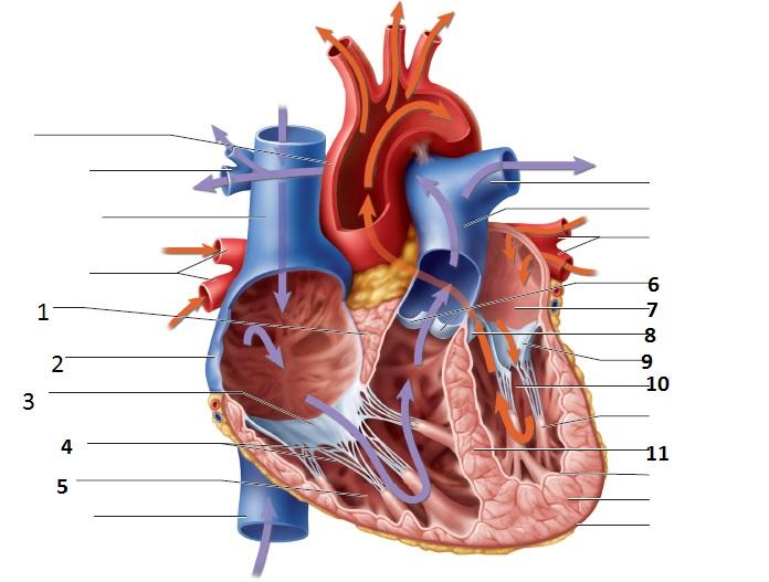
External structure of the heart with labels. 2. External features of the heart - SlideShare 2. THE HEART • The heart is a hollow muscular organ that is pyramidal in shape • It lies within the pericardium in the middle mediastinum • It is connected at its base to the great blood vessels. 3. General features of the heart • The heart has; • an apex and base • 2 surfaces; • Sternocostal surface • Diaphragmatic surface ... Ch. 19 Circulatory System- heart Flashcards - Quizlet Correctly label the external anatomy of the anterior heart. Place the labels in order denoting the flow of blood through the pulmonary circuit beginning with the right atrium and ending in the left atrioventricular valve. The first and last structures are given. Right atrium 1. tricuspid valve 2. right ventricle 3. pulmonary valve Human Heart - Anatomy, Functions and Facts about Heart All vertebrates, including humans, possess this type of circulation. The external structure of the heart has many blood vessels that form a network, with other major vessels emerging from within the structure. ... Label the Heart Diagram below: Practice your understanding of the heart structure. Drag and drop the correct labels to the boxes ... Solved Help Label the external anatomy on this posterior - Chegg Question: Help Label the external anatomy on this posterior view of a mammalian heart by clicking and dragging the labels to the correct location Coronary sinus Apex of heart Lert atrium Posterior interventricular branch of LCA Left pulmonary artery Left ventricle Left pulmonary veins Aortic arch This problem has been solved! See the answer
Correctly Label The Following External Anatomy Of The Anterior Heart ... The external anatomy of the human heart consists of the four chambers that form the apex of the heart. Each chamber has an apex that corresponds to a box. There are two boxes on each side of the heart: the atria and the ventricles. The left atrium is a branching organ. The pulmonary trunk contains the aorta and pulmonary veins. Solved Art-Labeling Activity: Overview of the external - Chegg art-labeling activity: overview of the external anatomy of the heart anterior view res great cardiac vein aortic arch right coronary artery left coronary artery left pulmonary veins ascending aorta left pulmonary artery anterior interventricular artery superior vena cava pulmonary trunk auricle of left atrium circumflex artery auricle of right … Surface projections of the heart: Borders and landmarks - Kenhub 1/4. The surface projections of the heart represent points on the thoracic wall that map out the outline and valves of the heart. These include four borders (superior, right, inferior, left) and four valves (left atrioventricular, right atrioventricular, aortic, pulmonary). The main reference points used for the surface projections of the heart ... Structure of the Heart | SEER Training The human heart is a four-chambered muscular organ, shaped and sized roughly like a man's closed fist with two-thirds of the mass to the left of midline. The heart is enclosed in a pericardial sac that is lined with the parietal layers of a serous membrane. The visceral layer of the serous membrane forms the epicardium. Layers of the Heart Wall
Labelling the heart — Science Learning Hub Blood transports oxygen and nutrients to the body. It is also involved in the removal of metabolic wastes. In this interactive, you can label parts of the human heart. Drag and drop the text labels onto the boxes next to the diagram. Selecting or hovering over a box will highlight each area in the diagram. Structure Of The Heart | A-Level Biology Revision Notes The two ventricles: these are the lower two chambers. They have thick, muscular walls which pump blood through the arteries. The heart is divided longitudinally into two sides by means of muscular walls. Each atrium is connected to its own ventricle through an opening which is guarded by a valve. Chapter 22 Heart Flashcards - Quizlet Label the coronary arteries in an anterior view of the heart. Label the order that blood flows through in the heart, using the arrows as guides. Label the components of the heart wall. Label the components of the heart as seen from a posterior view. Label the major coronary veins. Label the components of the conduction system. How to Draw the Internal Structure of the Heart (with Pictures) 1. To find a good diagram, go to Google Images, and type in "The Internal Structure of the Human Heart". Find an image that displays the entire heart, and click on it to enlarge it. 2. Find a piece of paper and something to draw with. Start with the pulmonary veins.
Structure and Function of the Heart
Internal Structure of the Heart | Contemporary Health Issues It is marked by the presence of four openings that allow blood to move from the atria into the ventricles and from the ventricles into the pulmonary trunk and aorta. Located in each of these openings between the atria and ventricles is a valve, a specialized structure that ensures one-way flow of blood.

mypicsainmarin: heart diagram with labels
Heart Anatomy: Heart Dissection - University of Washington The letters indicated in the text refer to the labels on the picture. The anterior surface of the heart is characterized by the presence of the large arteries leaving the base of the heart, the pulmonary trunk (H) and the aorta (G). The pulmonary trunk is the vessel that divides to give rise to the two pulmonary arteries going to each lung.

Lab 38: Structure of the Heart
Heart anatomy: Structure, valves, coronary vessels | Kenhub The heart is shaped as a quadrangular pyramid, and orientated as if the pyramid has fallen onto one of its sides so that its base faces the posterior thoracic wall, and its apex is pointed toward the anterior thoracic wall.
Heart - lab Flashcards | Easy Notecards
Heart Diagram with Labels and Detailed Explanation - BYJUS Diagram of Heart. The human heart is the most crucial organ of the human body. It pumps blood from the heart to different parts of the body and back to the heart. The most common heart attack symptoms or warning signs are chest pain, breathlessness, nausea, sweating etc. The diagram of heart is beneficial for Class 10 and 12 and is frequently ...

Pin on Heart Anatomy
Heart Anatomy: Labeled Diagram, Structures, Function, and Blood Flow Let's begin with the chambers of the heart. There are 4 chambers, labeled 1-4 on the diagram below. To help simplify things, we can convert the heart into a square. We will then divide that square into 4 different boxes which will represent the 4 chambers of the heart.
Virtual Squid Dissection
Heart Anatomy: size, location, coverings and layers : Anatomy & Physiology The heart wall is composed of three layers: the epicardium, myocardium, and endocardium. Location of the heart in the mediastinum. The superficial epicardium is the visceral layer of the serous pericardium. The middle layer is the myocardium and is composed mainly of cardiac muscle and forms the bulk of the heart.

Biology 156
A Labeled Diagram of the Human Heart You Really Need to See The human heart, comprises four chambers: right atrium, left atrium, right ventricle and left ventricle. The two upper chambers are called the left and the right atria, and the two lower chambers are known as the left and the right ventricles. The two atria and ventricles are separated from each other by a muscle wall called 'septum'.
Post a Comment for "44 external structure of the heart with labels"